Image

Interesting case of the week 7/1/2022 with Dr. Luke Watkins

This week’s interesting case is another great example of why imaging isn’t used to diagnose parathyroid disease or decide whether or not to do surgery. The patient is a 42 year old female who was noted to have elevated calcium levels for the past 4 years between 10.2 and 10.4. Her first PTH was checked only last year, and her levels have been from 64-90. These numbers are diagnostic for primary hyperparathyroidism.

After diagnosis, the next question is whether to pursue treatment. Any patient younger than 50 with hyperparathyroidism should be recommended for surgery. This patient found us and we met in clinic. We reviewed her blood work and discussed the surgery and recovery. On the day of surgery, her nuclear medicine scan was relatively unimpressive. Her ultrasound also did not see a tumor.

You can see the large left upper parathyroid adenoma that was removed that managed not to show up on the scan. In the hands of the wrong provider, a patient with these images might never make it to the operating room. Make sure you have your case evaluated by experts!

Sestamibi scanning is the preferred way to localize diseased parathyroid glands prior to an operation. HOWEVER, sestamibi scans are wrong at least 50% of the time.

35473c3 full-width


Image

Interesting case of the week 6/24/2022 with Dr. Luke Watkins

This week’s interesting case is about a 38-year-old male who we saw recently. He had elevated calcium levels noted incidentally on blood work at 11.3. His calcium was repeated (11.3 again) with a parathyroid hormone, which was 200, clearly showing hyperparathyroidism. We discussed the disease and recommendation for surgery based on his age and calcium levels.

On the day of surgery, his scan suggested two separate tumors, both on the right side. These scans usually show only one or no tumors. On the lower right image, you can see both the dark spot of the right lower tumor as well as the space between the two thyroid lobes being grayed out. This is usually seen with an upper gland tumor.

Both glands were over-functioning at close to the same level, although the upper gland is about half the size of the lower gland. This is why you want your surgeon to look at all four glands. Had we stopped after taking out the biggest signal, we would have only cured half of his disease. His post-operative parathyroid hormone decreased as expected, consistent with cure. He recovered well and had essentially no pain the night of surgery.

Make sure your surgeon knows what they are doing. See the experts!

b0246c3 full-width


Image

Interesting case of the week 6/17/2022 with Dr. Luke Watkins

This week’s interesting case highlights the need for an expert to evaluate your case.

The patient is a 65 year old male who had elevated calcium levels intermittently for 15 years. His other calcium levels were always in the upper limits of normal. Over the past two years, his calcium levels have maintained elevated between 10.3 and 10.8. His PTH was 29. His endocrinologist at a well known medical center told him he did not have the disease. He was having significant issues with sleeping and memory, and so he sent us his records to evaluate.

We discussed that his diagnosis was not definitive, but that there were not many other reasons to explain his elevated calcium levels and failure of his PTH to suppress. He elected to pursue surgery.

His scan on the day of surgery suggested a left upper parathyroid adenoma. An enlarged left upper gland was found at the time of surgery, as well as three normal glands. His tumor was removed and his post-operative PTH level decreased to 9. We treat parathyroid disease every day. Even endocrinologists at the best medical centers only see parathyroid disease some of the time.

Don’t waste your time with doctors who don’t understand parathyroid disease!

da30294 full-width


Image

Interesting case of the week 6/10/2022: Even people with small tumors or relatively low levels can have big symptoms, with Dr. Luke Watkins

This week’s interesting case is about a 71-year-old female who had been noted to have relatively high calcium levels on routine blood work. Her total calcium levels ranged from 9.5-10.0. Her ionized calcium levels were consistently above the upper limit of normal. Her PTH levels were checked multiple times, and ranged from 35-52, with an upper limit of normal. She had all of the symptoms typical of primary hyperparathyroidism, with fatigue, bone pain, insomnia, headaches, heartburn, brain fog, and irritability. She was noted on her most recent bone density scan to have osteopenia.

Lucky for her, her rheumatologist was not fooled, and picked up on the failure of her PTH to suppress with elevated ionized calcium levels. He referred her to us for evaluation. We reviewed her blood work, and discussed her options. She elected to proceed with surgery. Not surprisingly, her scans on the day of surgery were negative.

At surgery, it was noted that all four of her glands were abnormal and over-functioning. We performed a subtotal resection, leaving a portion of one of her glands behind. Her PTH levels decreased to 10, consistent with cure. She is another great example of how discordant symptoms can be with labs and tumor size. Even people with small tumors or relatively low levels can have big symptoms!

fe1bf3b full-width


Image

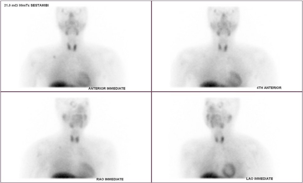
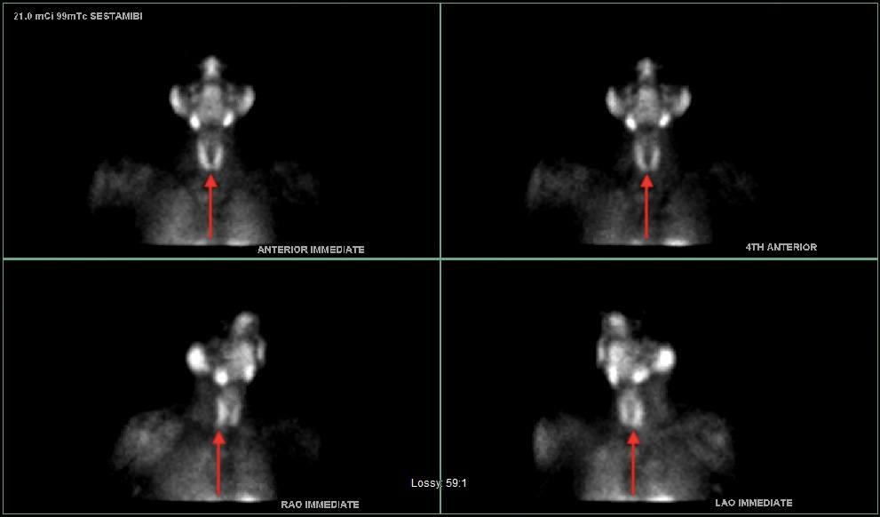
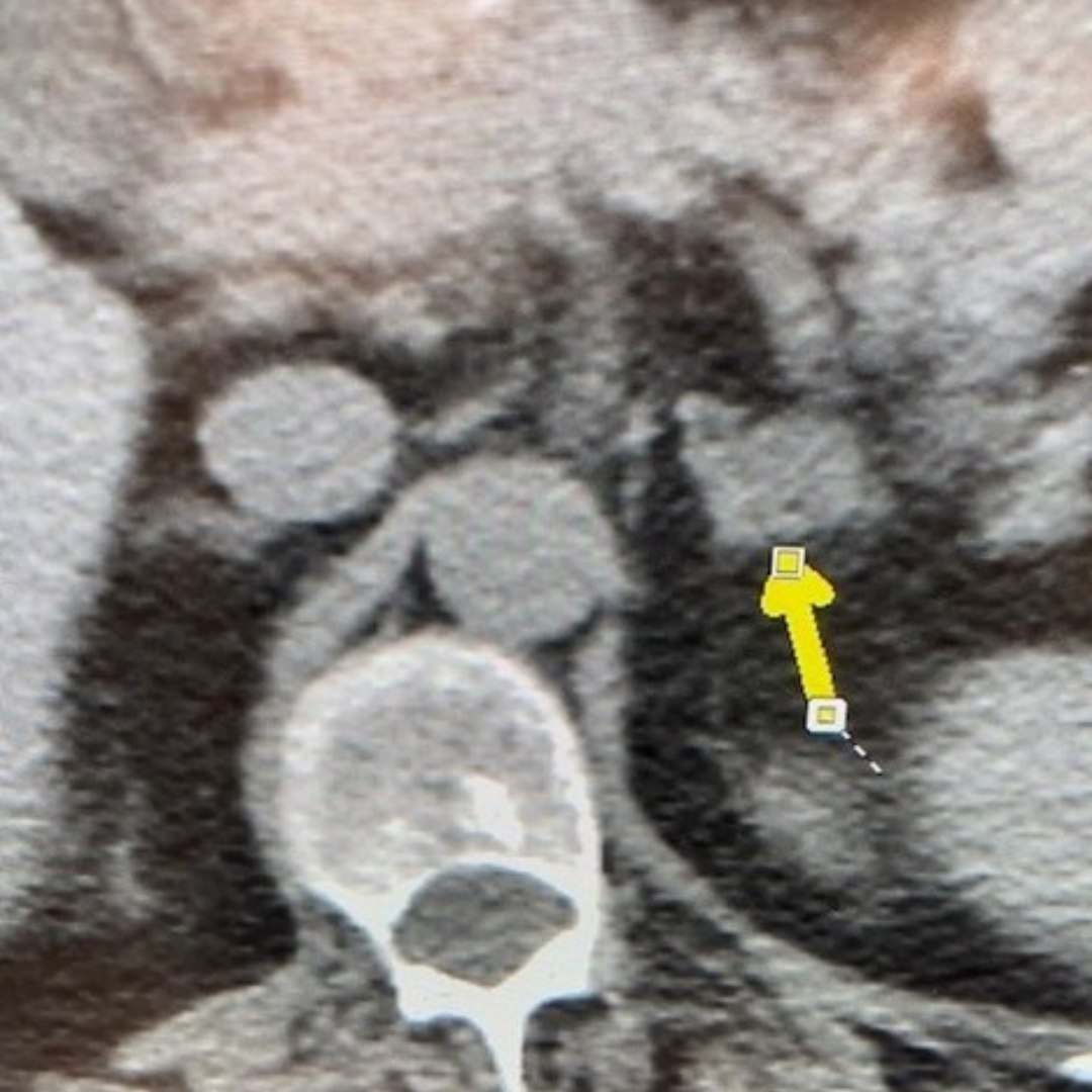
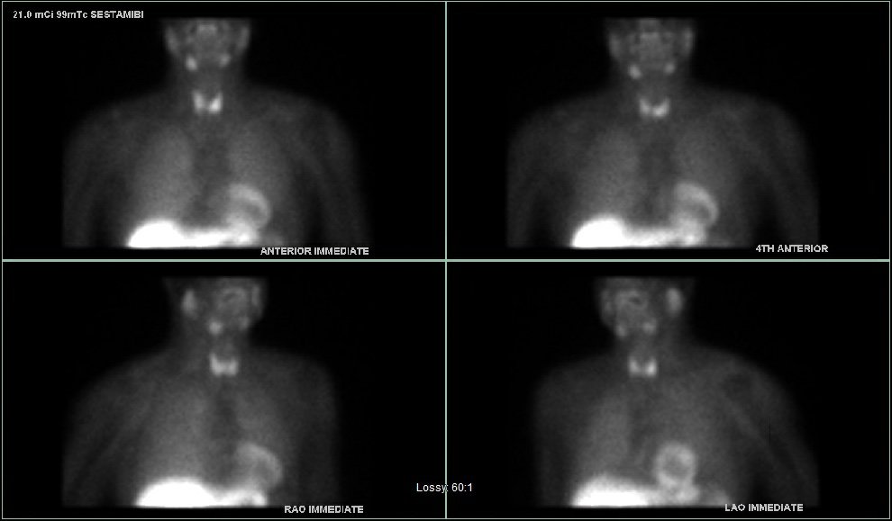
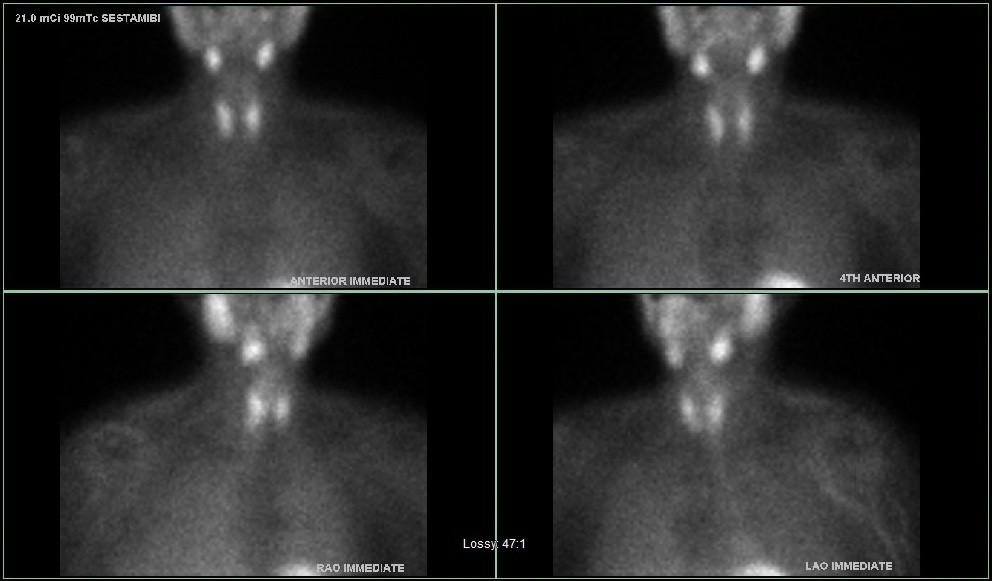
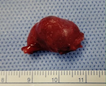
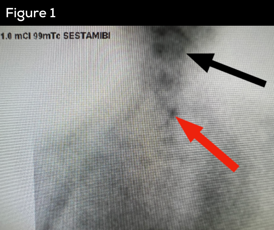
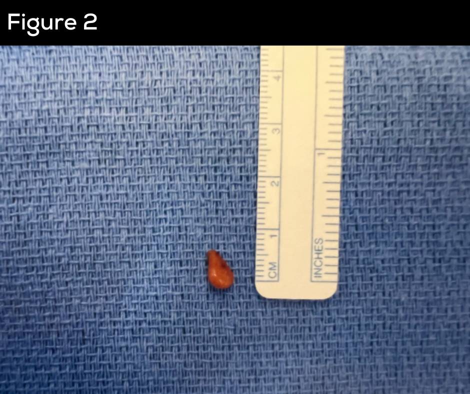
Interesting case of the week 6/3/2022: Parathyroid Tumor DEEP in the Chest with Dr. Drew Rhodes

A young, active, and otherwise healthy 30-year-old female was referred to us with primary hyperparathyroidism. Her day of surgery sestamibi scan at the Hospital for Endocrine Surgery is shown here.

Fortunately, the resolution of our new state-of-the-art machines incorporating our tried-and-true technology was able to see this parathyroid tumor in this young woman’s chest. The large arrow with the letters “SSN,” marks the suprasternal notch, or the top of the sternum, between the two collarbones. The smaller arrow is on the parathyroid tumor.

As the patient laid back on her pre-operative bed, the two marked lesions on the scan measured roughly 7 centimeters from one another, or almost 3 inches! The parathyroid tumor was actually closer to the heart (the bright structure on the lower right of the image) than the neck.

At most outside hospitals, a parathyroid tumor this deep in the chest would warrant a consultation with a chest surgeon and the discussion of undergoing a median sternotomy (bone saw cutting the chest cavity open to allow the surgeon to hopefully find the gland causing the primary hyperparathyroidism). But at this Norman Parathyroid Center, this patient underwent our standard neck incision. We slowly and carefully pulled the tissue up from the chest, within which the parathyroid gland was hiding.

The parathyroid tumor was found and removed allowing this young and healthy patient to get on with her life, without the scary proposition of having to heal for months from her chest having been cracked open!


Image

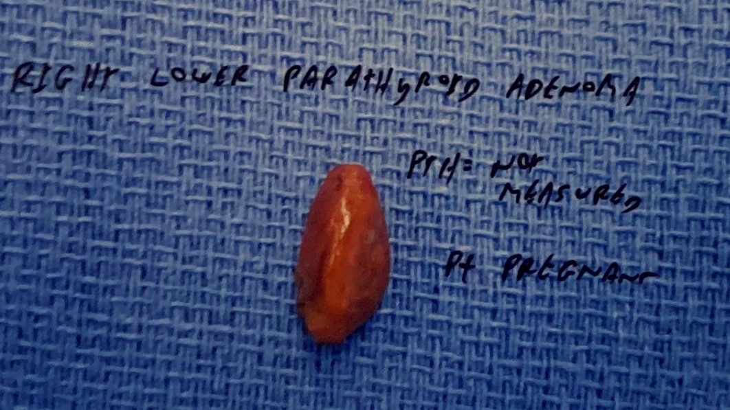
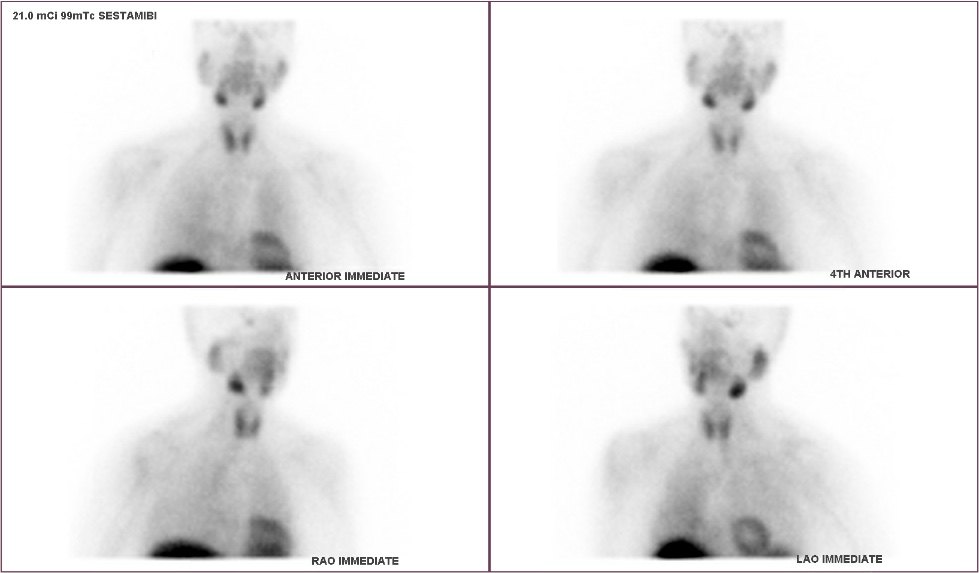
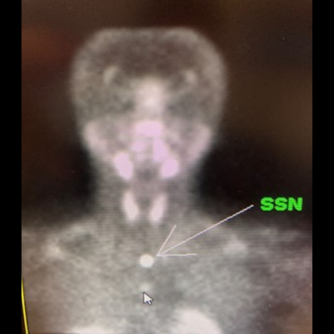
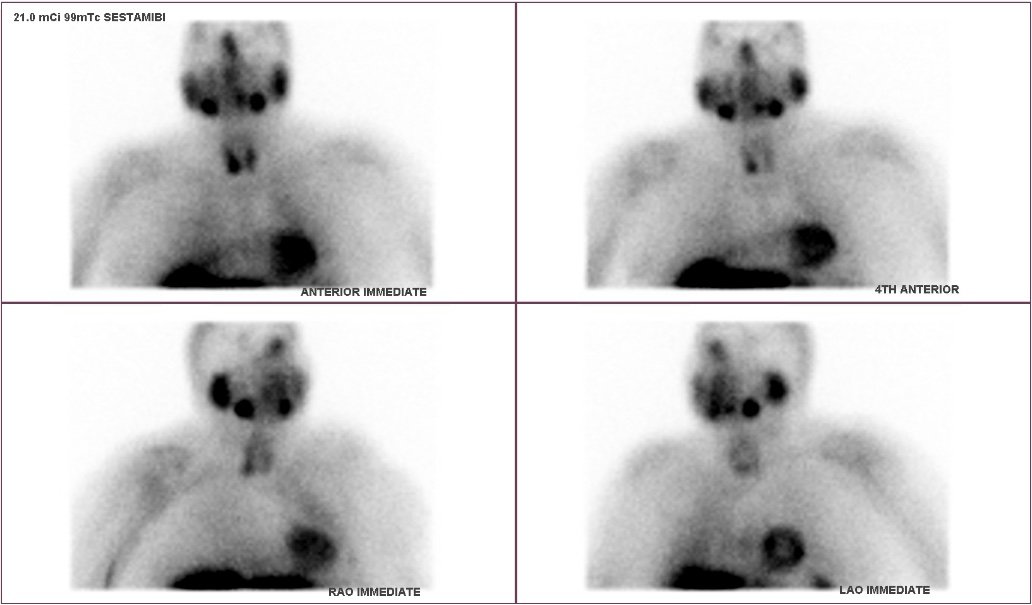
Interesting case of the week 5/27/2022: The Norman-Way in Pregnancy with Dr. Drew Rhodes

A 33-year-old pregnant female, towards the end of her 1st trimester, presented to us with long-standing hypercalcemia. We know that hypercalcemia (especially in the pregnant mother) needs to be fixed as soon as recognized. The best time to do this surgically in pregnancy is early in the 2nd trimester.

Our young mother had hypercalcemia for at least the last 8 years! She suffered miscarriages, first in 2014, but fortunately had two viable pregnancies over the last three years. However, she suffered with preeclampsia and preterm labor in both, requiring urgent caesarian section.

In preeclampsia, the mom has high blood pressure and high protein levels in her urine. This can lead to generalized swelling and abdominal pain, vision changes, and debilitating headaches, even stroke.

High calcium in the pregnant mother can not only cause preeclampsia, but problems for the unborn fetus, such as stunting of growth, preterm labor and delivery, and too low blood calcium levels, even tetany and death.

Fortunately, she was referred to us. We have a protocol in-place to intervene EARLY in the 2nd trimester to hopefully prevent the dangerous effects of hypercalcemia.

Fetal heart tones were obtained by our obstetric provider on the morning of her surgery. She was the 1st case of the day. Anesthetic modifications were made, and an ultrasound only (performed by her surgical team) is conducted (NO SESTAMIBI SCAN, NO injection of the radiotracer).

Her surgery was quick, less than 15 minutes, where a large adenoma was found and excised. Her vital signs and fetal heart tones were stable throughout the procedure and in the recovery room, where her post-operative blood parathyroid hormone (PTH) level was 8.5, from as high as 62 preoperatively (signifying CURE)! She went home later that morning knowing she made a LIFE-CHANGING decision.


Image

Interesting case of the week 5/20/2022 with Dr. Luke Watkins

This week’s interesting case is about a 62-year-old female who was recently noted to have elevated calcium levels. She had not been to the doctor for awhile. Her routine blood work noted calcium levels from 11.6 to 12.1. Her PTH was checked twice and was 241 and 460! With numbers like these, her diagnosis was clear. Still, it took her six months to to get referred to see us. She didn’t notice many symptoms, but was being treated for osteoporosis.

Prior to surgery, she had a nuclear medicine scan that was read as non-localizing. On the da of surgery, her scan clearly showed a right upper parathyroid adenoma. Her surgery was about 25 minutes, her tumor was removed, and her other glands were confirmed to be normal.

We often point out how small numbers can still cause big symptoms. Here is an example from the other end of the spectrum: BIG numbers, but almost no symptoms whatsoever. Her osteoporosis, however, would not get better without parathyroid surgery.

8798957 full-width


Image

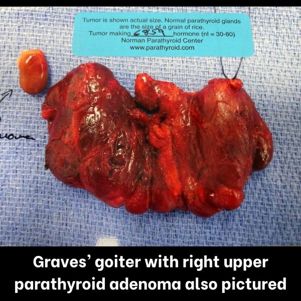
Interesting case of the week 5/13/2022: Two Birds, One Surgery with Dr. Drew Rhodes

Whether recognized during your initial consultation, or during your day-of surgery preoperative evaluation, your thyroid CAN be dealt with at the time of your parathyroid surgery. We have two examples to illustrate this below.

A 59-year-old female patient came to us with elevated calcium levels for the last two years. During our detailed evaluation, we discovered she had a multinodular thyroid with several large nodules that had “biopsied benign.” Further, she had been on methimazole, a drug used to dampen the effects of a hyperfunctioning thyroid nodule.

Typically, this drug is used for 12-18 months to see if the hyperfunction can be reversed. As in this patient, it oftentimes DOES NOT and surgery is required to remove the portion of the thyroid not working properly. This patient was found to have her entire thyroid enlarged, requiring a total thyroidectomy at the same time of her parathyroidectomy.

For our second case, a 65-year-old female patient came to us with long-standing high calcium levels as evidenced by her blood work, osteoporosis, and 20-year history of kidney stones.

She also had a similar lumpy/bumpy thyroid as the 1st case example, which was being “watched” by her home providers. She had not had a recent thyroid ultrasound or nodule biopsy, all of which previously showed a “big” thyroid with benign nodules. During our thorough preoperative evaluation, you could see the contour of her big thyroid protruding along the front of her neck. When asked about swallowing or breathing difficulties or hoarseness (all potential symptoms of an enlarged thyroid gland), she confirmed.

Both “dual” cases (thyroid and parathyroid excisions) were completed safely in under an hour, allowing the patients to sleep in their own beds that evening (or return to one of our partner hotels). These patients are forever grateful for not having to undergo two separate surgeries, proving (once again) the “Norman-Way” is the ONLY way!

7a2f8c2 full-width


Image

Interesting case of the week 5/6/2022: Importance of the Norman-Way in Finding YOUR Tumor with Dr. Drew Rhodes

A 72-year-old gentleman visited us from across the state, suffering with hypercalcemia over the past year, contributing to overwhelming fatigue, constipation, brain fog, and kidney stones.

We cannot emphasize enough the importance of you having your scans on the day of surgery and having the images interpreted by YOUR surgeons.

The quick 15-minute, day of surgery sestamibi scan allows us to not only best understand where a hyperactive parathyroid tumor may be hiding, but also provides the injection of the rapidly decaying radiotracer used intraoperatively to understand which

In this gentleman’s case, as you can see from the subtle protrusion laterally from the midline on the left lower image (white arrow), he had a deep and descended LEFT UPPER tumor. As the patient rotated his head to the right, the deep left upper adenoma shifted laterally, signaling a rare location for an upper gland, which had descended (settled low in the neck during in utero development). Typically, an upper gland is along the upper pole of the thyroid gland.

9 out of 10 radiologists (more likely 10 out of 10!), who typically interpret these pictures for the surgeons at other hospitals (NOT at The Hospital for Endocrine Surgery), would deem this tumor a LEFT LOWER adenoma. The surgeon at the other hospital would be hell bent to find an abnormal LOWER tumor and would look-and-look in the typical lower positions, and ultimately, be unsuccessful (The patient WOULD NOT be cured).

Fortunately, this patient had the Norman-Way and was cured in less than 30 minutes, able to achieve the quality of life he deserves!

11deb5f full-width


Image

Interesting case of the week 4/29/2022 with Dr. Luke Watkins

Today we have a 29-year-old male who was referred to us after he was noted to have significantly elevated calcium levels. He had noted symptoms of fatigue, brain fog, irritability, and problems sleeping - all hallmarks of parathyroid disease (hyperparathyroidism). Read more about the symptoms of parathyroid disease here: https://www.parathyroid.com/parathyroid-symptoms.htm

He was noted to have a calcium level of 12.2 and a parathyroid hormone level of 381.8. Given his age and calcium level, he was recommended for surgery. Learn more about parathyroid surgery here: https://www.parathyroid.com/treatment-surgery.htm

His scans on the day of surgery strongly suggested a left upper adenoma, which is what was found at surgery. He recovered well with a post-operative PTH level of 17, clearly indicating cure.

Although our average patient is in their 60s, hyperparathyroidism can affect people of any age. Younger people typically have more pronounced symptoms and larger tumors. This patient got on the path to cure when his primary care doctor noted a significantly elevated calcium. Don’t let your symptoms go undiagnosed or untreated!

410fe90 full-width


Image

Interesting case of the week 4/22/2022 with Dr. Luke Watkins

This week’s case is about a lovely 65-year-old woman who came to see me. She had an automobile accident in 2016 which required a tracheostomy (a breathing tube placed directly through the neck) to be placed. Thankfully, she recovered from her injuries. She was noted during this process to have elevated calcium levels, which have been present for the last ten years.

Recently, she had calcium and parathyroid hormone levels checked, which showed an elevated calcium and PTH. We talked and reviewed her case. She had significant osteoporosis as well as issues with fatigue, memory, concentration, and irritability. Given her osteoporosis, I recommended surgery.

On the day of surgery, her scan suggested a left upper adenoma. She had significant scar tissue from her tracheostomy that required extra care be taken. The tumor was a left upper adenoma. He post-operative PTH level decreased appropriately consistent with cure. She did great after surgery. We were able to free some of her scar tissue from her old scar to improve her cosmetic result.

Previous surgery can increase the risk of nerve injury by a factor of ten, however our patient was cured without complication because she selected a team of experts.

21768bb full-width


Image

Interesting case of the week 4/15/2022 with Dr. Luke Watkins

This week’s interesting case is about a 32-year-old male from Missouri with a significant medical history. When he was 6, he was diagnosed with chronic lymphocytic leukemia. He underwent a bone marrow transplant and did very well from his treatment. Unfortunately, he developed an osteosarcoma that required an amputation of one of his legs. Again, he did well from this.

He was having significant fatigue, and he was noted to have an elevated calcium during his work up, and follow up testing confirmed an elevated calcium and parathyroid hormone. He did not live far from an academic medical center, however his aunt also had parathyroid disease and was treated by us. He elected to pursue treatment with us as well.

On the day of surgery, his scan faintly suggested a tumor in the left upper location. The left upper adenoma that was noted at surgery can be seen in the photo here. His parathyroid hormone decreased after surgery, consistent with cure.

Given his history, the question that concerned him most should not come as a surprise: Was it cancer? His pathology showed only benign parathyroid glands, which is almost always the case.

Every patient has a different story before they come to us, but they almost always end the same way: cure!

c6d4ef6 full-width


Image

Interesting case of the week 4/8/2022 with Dr. Luke Watkins

This week’s interesting case is about a 69-year-old female who had primary hyperparathyroidism for three years. She had osteopenia and multiple symptoms of hyperparathyroidism, including low energy, bone pain, memory and concentration issues, and irritability. After being diagnosed, she was referred to our clinic for evaluation.

She elected to pursue surgery with us. On the day of surgery, her imaging noted a somewhat interesting finding. The arrow denotes the finding. Most findings on nuclear medicine scan are off to the left or the right, but this one is in the middle when the picture is taken from straight ahead. When the camera is moved to either side, the lesion moves to the opposite side.

This type of movement is typical of lesions located behind the esophagus. It is why we do our imaging this way, Many places do not take pictures from the sides, which means this tumor likely would have been missed. Even worse, some surgeons may have decided not to operate because the tumor was not seen (thinking this was part of the thyroid).

Her surgery was uneventful and her post-operative parathyroid hormone level decreased consistent with cure. She felt great after surgery, as so many of our patients do. We see these types of “complicated” cases every day. Trust the experts!

7af447f full-width


Image

Interesting case of the week 4/1/2022: Two hormonal dysfunctions, one patient with Dr. Drew Rhodes

This week, we saw a 68-year-old female from WV who came to us with long-standing hypertension, kidney decline, and typical symptoms seen with primary hyperparathyroidism (PHPT), including bone pain, fatigue, reflux, muscle weakness, and brain fog.

In speaking with her on initial consultation, we saw her biochemical dysfunction as seen in the photo, blood calcium as high as 11.4 and corresponding PTH 173, with poor kidney function.

In speaking with her further about her medical history, she recounted several decades of hypertension. She also mentioned a significant finding in her medical history, hyperaldosteronism. Elevated aldosterone (salt) levels can contribute to elevated blood pressure and kidney decline. 20% of patients with resistant hypertension (high blood pressure treated with medicines) can have primary aldosteronism caused by a benign (non-cancerous) adrenal adenoma (tumor).

A less than 30-minute operation with the Carling Adrenal Center (our sister surgeons at the Hospital for Endocrine Surgery) can cure this patient’s long-standing high blood pressure. Fixing two chronic problems at one surgery center of excellence, what can be better?

The right lower parathyroid tumor (as seen in the photo) was the only gland requiring removal. The PTH level in the recovery room was 15, signifying intraoperative cure.

With the Carling Adrenal Center as our neighbor, the patient was able to have confirmatory blood work and an adrenal protocol CT scan after her parathyroid surgery and before heading home. The adrenal CT scan showed a 1-2 cm tumor in the left adrenal gland, the culprit of her long-standing high blood pressure! We were able to send this information back with the patient to share with her kidney doctor, showing help was on the way!

She will be heading back our way in the near future, as she said, “to escape the cold and finally fix what has been hurting me all these years, another trip to Tampa sounds perfect!”

c08d657 full-width


Image

Interesting Case of the Week 3/25/22 with Dr. Luke Watkins

This week's interesting case is about a 68-year-old male with hyperparathyroidism. He had been noted to have high calcium levels for three years. He had been suffering from significant fatigue. During this time, he had a heart attack, and his kidney function had worsened. His calcium and PTH levels were just barely outside the normal range.

When we discussed his case, we discussed his lab work and how, despite being relatively normal, clearly demonstrated hyperparathyroidism. We discussed how even with relatively “low” calcium and PTH levels, people can still feel significant symptoms. Most importantly, we discussed his declining kidney function. While there are more common causes of kidney dysfunction, hyperparathyroidism is curable where many others are not.

On the day of surgery, his scan suggested a left upper tumor. As you can see in the picture (and as we have seen so many other times... imaging can be misleading!), he also had a right upper adenoma found while evaluating all four glands.

He did great post-operatively with a significant drop in his PTH indicating cure. His case is a great example of the significant effects hyperparathyroidism can have even with small numbers.


Image

Interesting Case of the Week 3/18/22: Misleading preoperative imaging with Dr. Drew Rhodes

One of our patients from last week visited us from Houston, TX. She had clear biochemical evidence of primary hyperparathyroidism for 3 years, blood calcium was as high as 10.6 with corresponding PTH 120 and Vitamin D 13. She felt lousy. She had bone pain, fatigue, difficulty sleeping, headaches, heartburn, and brain fog. She and her husband wanted to find her old self again. She had an ultrasound at her home doctor’s office suggesting a right middle-thyroid cystic thyroid nodule. This finding led to more questions than answers. She wondered if she had a thyroid nodule that needed intervention. Her home doctor suggested needle-aspiration of the nodule to see if they could obtain more information.

We took one look at the nodule with our standard day-of-surgery ultrasound and confirmed the nodule to be a mixed cystic and solid parathyroid gland, not a thyroid nodule. The patient did not need expensive imaging tests at home, but rather the Norman way in a bad way.

The excised glands can be seen in the picture below. The large mixed cystic and solid tumor in the right lower position, significantly over-producing PTH as evidenced by the 10,015 unit number (normal intraoperative production approximately 30-80 units), along with another tumor, in the left upper position, which would have prevented cure had it not been found and removed.

The scans don’t find the tumors. Surgeons find the tumors, and then make appropriate decisions based on what is found to best help our patients. Her post-op PTH was 17.1 (from 120 pre-op) in the recovery room signifying cure. When I spoke to her that evening, she was already feeling better than she had in years! This is why we do what we do!


Image

Interesting case of the week 3/11/22 with Dr. Kevin Parrack

Today’s interesting case is a great example of how much patience is required of some patients to finally get cured after having a bad experience with their first surgery and getting mixed messages afterwards.

We operated on a nice lady who had parathyroid surgery close to ten years ago at a major university hospital. During that surgery, one of her parathyroid glands was taken out and her right thyroid lobe was removed, but none of the other three parathyroid glands were found despite an extensive operation that lasted eight hours. Unfortunately, her calcium and PTH levels remained elevated after the surgery. As it would for anyone, such an experience left her shaken.

Before and after this operation she was suffering from classic parathyroid symptoms. She had fatigue, brain fog, palpitations and chronic pain. As bad as she felt from parathyroid disease, she told me that she was afraid of having another surgery. Compounding her worries was the fact that she had years of negative imaging. Sestamibi scans, ultrasounds and 4D CT’s had all been unsuccessful in localizing her parathyroid tumor.

Before her first operation her PTH was over 100, but by the time she contacted us, her PTH was over 250. She was so tired of feeling bad that she was willing to consider taking the risk of another surgery. Although I of course had to explain to her the risks inherent in re-operative surgery, I also encouraged her that she is not alone. Many people have been through bad experiences with their first surgeries, and had frustrating negative imaging for years thereafter, but there is still hope to be cured. It is worth it to try because this disease is a difficult burden to bear, and the joy of being cured and the symptoms going away makes all the struggle worth it.

I also perhaps immodestly explained that although complications and difficulties can happen with any surgery, we have focused our careers on this disease, including the difficult cases, to make those risks as low as possible. I expect even difficult re-operative cases to recover well.

Many patients who are not cured by surgery and are referred to us from other places have upper parathyroid tumors in the deep space of the neck, behind the thyroid, on the spine, and the previous surgeon was uncomfortable looking back in this area.

Figure 1 shows what I saw during her ultrasound. The red arrow points to the thyroid. The blue arrow points to a dark spot that is either a parathyroid adenoma, or a lymph node.

Needless to say I was excited. A dark spot behind the left thyroid lobe perfectly fit my suspicion of a parathyroid tumor in this area. Of note, the ultrasound showed no thyroid lobe or parathyroid tumor on the right.

I will shamefully admit that I thought to myself that this would probably be an easy re-op case with a nice parathyroid tumor in such an obvious location. Well, in the operating room that spot behind the left thyroid lobe turned out to be a benign lymph node, not a parathyroid tumor. We looked in the correct locations in the left neck for upper and lower parathyroid tumors and none was found. With that done it was now time to look at the right side of the neck.

Usually when someone has no thyroid lobe on one side, big parathyroid tumors on that side are obvious on imaging. She of course had multiple negative imaging studies so one would think the odds of a parathyroid tumor on that side would be low, but our sestamibi scan told a different story.

In figure 2, the orange arrow points to the left thyroid lobe. It looks like there is nothing on the right side. Figure 3 shows another angle with the patient's head turned. The arrow points towards a finding that might be a parathyroid adenoma in the deep space of the neck.

In the operating room we carefully exposed the important structures in the neck, and sure enough, on the right side, behind the right recurrent laryngeal nerve, sitting on the spine was a deep right upper parathyroid adenoma.

There was a lot of scar tissue in her right neck from the previous surgery and it densely involved the right recurrent laryngeal nerve that was in front of this tumor. But once we got past this important nerve and got to the tumor, it was clear the the previous surgery had not extended back to the bulk of the adenoma.

Her PTH in the recovery room dropped down to 19 (from over 250 before the surgery). I had such a good time speaking to her the evening after her surgery because not only was she excited to put all this behind her, she told me that the pain which had been plaguing her for years was just gone.

She asked me to share her story because she wanted to help other people who are going through what she just got past. I hope those of you who read this and are dealing with the disease, particularly those who were not cured with surgery, or even had a complication from a surgery, can be encouraged that there is hope!


Image

Interesting case of the week 3/4/22 with Dr. Luke Watkins

This week’s interesting case is about a 68-year-old female who came to see us from Idaho. Her calcium had been monitored for 5 years, and her PTH had been checked multiple times during that span. Her calcium ranged from 9.6 to 10.6, with PTH ranging from 46 to 94. Despite this, she was treated with vitamin D (which is used to treat low calcium and help with mild bone loss, but is useless with high calcium). She was noted to have osteoporosis and several typical symptoms of hyperparathyroidism.

She found us online and decided to trust us instead of her local surgeon. As expected, her imaging was non-localizing. Her surgery was uneventful, with a small but enlarged right lower parathyroid gland identified, with otherwise normal glands. Post-operative PTH decreased as expected.

This patient has the same story we hear over and over again: failure to correctly diagnose when parathyroid disease is suspected or recommend intervention when it is diagnosed. However, when she took control of her care, she travelled across the country to see us. For her, getting the right diagnosis and treatment was worth it.


Image

Interesting case of the week 2/25/22 with Dr. Luke Watkins

This week's interesting case is a great example of how imaging can be wrong. An 84-year-old retired physician was sent to us by his kidney doctor. He was noted to have primary hyperparathyroidism as a cause for his worsening kidney function, and the only cause that could be cured. He had his thyroid taken out 50 years ago and was in good health otherwise.

His nuclear medicine scan on the day of surgery shows vague signal where the thyroid used to be. Nothing significant was seen on his ultrasound, no remaining thyroid tissue or parathyroid glands. We found four enlarged glands in their typical locations, and performed a subtotal parathyroidectomy. After surgery, his parathyroid hormone decreased appropriately consistent with cure. He recovered well from surgery.

The important aspect of this case (other than cure without complications) is that this patient’s scans did not identify a tumor. His ultrasound was unable to see the glands even without a thyroid in the way. His nuclear medicine scan did not localize to a tumor. In his case, we had a high suspicion that he might have four glands involved based on the fact that there was some signal on both sides.

Consider a patient with the same disease who still has their thyroid. What if their doctor ordered the scan to diagnose them or decide whether or not they needed surgery? A patient with a curable disease would never make it to surgery, simply because scans can be, and frequently are, wrong.


Image

Interesting case of the week 2/18/22: Modest PTH can be hiding monster tumors, with Dr. Drew Rhodes

This week, we had the pleasure of treating a 70-year-old gentleman from Washington.

He was taking medication for hypertension and atrial fibrillation, both of which can be contributed to by elevated blood calcium levels. He endorsed typical symptoms of hypercalcemia, namely bone pain, fatigue, and insomnia, along with typical neurocognitive complaints such as memory and concentration difficulties and irritability.

He needed the Norman-Way in a bad way. Unfortunately, despite his elevated calcium levels (above 10.1 in an adult twice by July 2020), when his provider checked his PTH and found it to be “normal” (less than 65 in the lab listed below), this did not trigger a referral to us.

Based on past cases we’ve told you about, we know that with a blood calcium level of 10.6, “normal” physiology would be if the PTH was 20 or less. A PTH level of 50, as in this case, is 2.5 times higher than what is really normal! You can find his pre-operative laboratory values in the photos above.

Fortunately, the patient was his own best advocate. He was sick-and-tired of being sick-and-tired and decided to reach out to us in late 2021, after his providers wanted even more blood work and weren’t quite ready to pull the trigger on a surgery consult to get him cured.

Despite his modest PTH levels, we found two monster tumors in his neck (see photo above). Each tumor was putting out significant levels of PTH hormone, 2823 units from the right upper parathyroid adenoma and 3313 units from the left upper gland (normal intraoperative production should be around 50 units). His lower glands were clinically normal and biopsied.

His post-operative PTH in the recovery room was 20 (finally normal)! Our patient recovered nicely and is on his way to feeling like a new man.


Image

Interesting case of the week 2/11/22 with Dr. Luke Watkins

This week's interesting case is about a 69-year-old female who had elevated calcium levels for ten years. She had relatively few symptoms, but a bone density scan showed she had osteopenia (just short of osteoporosis). The bone loss was in her wrist more than the weight bearing joints, which is typical of parathyroid disease.

Over the last few years, she had her parathyroid hormone checked, which ranged from 26 to 48. Her parathyroid hormone levels were not suppressed with her elevated calcium. Given her bone loss, she wanted to pursue treatment.

On the day of surgery, her scan weakly suggested a right upper gland as the source of her disease. An enlarged gland was found at surgery. Her PTH level dropped post-operatively showing she was cured.

Any physicians would see her “normal” PTH and plan for observation while her osteoporosis worsened. This demonstrates the importance of getting evaluated by experts.

The Norman Parathyroid Center is the leading parathyroid treatment center in the world, performing about 10% of all parathyroid surgery in the US. We are located in Tampa, Florida where hyperparathyroidism is the only disease we treat. We perform nearly 4,000 parathyroid operations annually with a cure rate over 99% via a minimally invasive operation that typically lasts about 20 minutes and evaluates all four parathyroid glands.


Image

Interesting Case of the Week 2/4/22: Patients with modest parathyroid hormone levels can have large parathyroid tumors

Mr. B is a 39-year-old police officer who presented with muscle and joint pain that began a year ago. This prompted an evaluation that uncovered a serum calcium level of 10.1 mg/dL.

Subsequent lab testing revealed a parathyroid hormone level of 71 pg/mL (reference range 15-65 pg/mL), low vitamin D levels, and a high level of calcium in his urine (634 mg/day).

Primary hyperparathyroidism (pHPT) was diagnosed based upon these findings. The consequences of living with pHPT include loss of bone strength, kidney problems (a high risk of kidney stones and loss of kidney function), and a higher risk of heart attack and stroke.

Furthermore, pHPT is associated with a wide range of symptoms that can worsen quality of life. Mr. B reported several of these symptoms, including fatigue, problems with mental focus, and difficulties with memory.

Mr. B was taken to the operating room, and all four of his parathyroid glands were identified and tested for function. One very large parathyroid tumor was found and removed (pictured here).

Although patients with modest calcium and parathyroid hormone levels tend to have modest-sized parathyroid tumors, Mr. B’s parathyroid tumor was remarkably large. A normal parathyroid gland is approximately the size of an uncooked grain of rice. Although Mr. B’s parathyroid hormone level was only 9% over the reference range, his parathyroid tumor was more than 200 times the volume of an average parathyroid gland.

Parathyroid patients from all over the world travel to Tampa for their parathyroid surgery. We have performed over 60,000 parathyroid operations, more than the top 20 US universities combined.


Image

Interesting case of the week 1/28/22 with Dr. Kevin Parrack

Our first case involves a lovely and very well organized lady in her 70’s who had labs suggestive of long-term primary hyperparathyroidism. Both her calcium and PTH levels were high, and her calcium levels were above 11 repeatedly, all the way up to 12.1. That is concerning. She had several of the typical problems that plague parathyroid patients, most bothersome were her brain fog, heart palpitations and hypertension.

What made her case special, other than the long time she had likely been dealing with this problem, was that her thyroid had been removed for cancer in the past. This is a pretty common reason why patients are referred to our center. If they had neck surgery in the past, they will have scar tissue around their parathyroids and it is even possible that some of their parathyroids were removed during their previous surgery.

This can be a real problem because if three parathyroid glands were accidentally removed or killed during the previous surgery, and in our following surgery we remove their tumor, which happens to be their last parathyroid gland, then their PTH level will drop to zero, a condition known as permanent hypoparathyroidism, which is frustrating because to treat it patients take calcium supplements repeatedly every few hours for the rest of their lives.

Now most surgeons who remove a thyroid are experienced enough to reduce the odds of killing multiple parathyroid glands down to a very low level. So it’s a risk we take having to follow in another surgeon’s footsteps, but usually a low risk.

For our nice lady, there were a few additional factors. Her thyroid cancer had spread at the time of her past surgery, and therefore she had removal of the lateral lymph nodes in her neck on both sides. In addition her surgery was done in the 1960’s and they did what is known as a radical neck dissection, which is a more disruptive surgery than what is typically done today.

So we had a long talk that although most thyroid surgeons are successful at not removing multiple parathyroid glands during a thyroidectomy, she had an extensive operation and the odds in her case were higher than in a standard, modern thyroid surgery.

We had a detailed conversation about the options of removing her tumor completely with cryopreservation as an insurance policy, vs subtotal parathyroidectomy while keeping a total parathyroidectomy with autotransplantation as a back up plan if the subtotal was not technically feasible.

After having this detailed conversation she came to see us in Tampa and she brought with her the operative report and pathology note from the 1960’s!!! I often do between one and four re- operative cases a day at our center, and this is the only time I can ever remember a patient successfully tracking down an op note and path report from over 50 years ago. It had been pulled off a microfiche (remember those?), was a bit faded and copied on pink “receipt” paper (remember that?). But the most important part of the documentation as far as I was concerned was the path report that clearly stated three parathyroid glands had been found in the operative specimens. This meant that three of her four parathyroid glands had been removed during her thyroid surgery.

I was stunned and impressed that the patient had tracked down medical reports from so long ago, and further impressed that the pathologist had been so careful that he found three individual parathyroid glands in a sea of thyroid tissue, lymph nodes, fat and cancer. Frankly that is outstanding work.

It also clarified our patient’s optimal surgical plan. Removing her tumor completely would be an inferior option for her since the risk of permanent hypoparathyroidism would be unacceptably high even if we used cryopreservation or autotransplantation. So our goal was a subtotal parathyroidectomy.

Figure 1 shows the sestamibi scan.The red arrow points to her parathyroid tumor. The black arrow points to one of her submandibular glads, which is just under her jaw, to give you some reference. In the operating room her scar tissue had thankfully softened up quite a bit over the years and we were able to safely approach her parathyroid tumor.

Figure 2 shows what we took out.

Turns out the tumor wasn’t very big; we assume this may be related to the scar tissue restricting its growth, but you never know. If you look carefully at the picture, you’ll see the top part of the tumor, closest to the ruler, has a straight cut edge. That is because when we did the surgery I carefully cut through the tumor, leaving a piece in her neck, that is roughly the size of a normal parathyroid gland, on its original blood supply. This little piece will make enough hormone so that her calcium won’t drop too low.

I also marked the location of this “remnant” parathyroid with a blue stitch so that if this little tumor fragment grows larger (usually takes 15 years or so if it’s going to be a problem), I can always find it again in the future and trim it down.

Thankfully after surgery her PTH came down, but not too low. I am so impressed with her efforts to get her records and the outstanding work by the pathologist. These two things helped me pick the right surgery for this very special, well organized lady.


Image

Interesting case of the week 1/21/22: 25 years of high calcium with Dr. Luke Watkins

Our interesting case this week is about a 69-year old-female who had elevated calcium levels for 25 years before coming to see us. Over that span, she had developed atrial fibrillation, high blood pressure, and even had a stroke. Despite all of this, she kept working. Recently, her fatigue had increased significantly, making getting through the day difficult. Her most recent blood work showed her calcium and parathyroid hormone to be elevated at 10.5 and 111, respectively.

She decided to come see us at our COVID-free hospital. Her scan that morning showed an obvious parathyroid lesion below the thyroid.

What is interesting is that this gland, despite the fact that it is lower than the other parathyroid on the side, is actually the right upper parathyroid gland.

At surgery, we found her tumor as expected, very far back in her neck, as well as three other normal glands. Her parathyroid hormone levels dropped significantly. She did great after surgery, and had minimal complaints.

Parathyroid glands in this location are very frequently missed by inexperienced surgeons. Most scans that are done elsewhere are read by the radiologist and/or the surgeon as being a lower gland. We know how scans are useful and misleading. Come see the experts!


Image

Interesting Case of the Week 1/14/22: Another case against focused parathyroidectomy with Dr. Drew Rhodes

A 76-year-old patient from Santa Fe had a specialized CT scan suggesting a 1 cm left upper parathyroid tumor. She then visited with an area surgeon who suggested a “focused” surgery to go find the gland seen on imaging and then check intraoperative PTH levels to watch the decline in the level from the preoperative value, which was as high as 107, with blood calcium levels as high as 11.4.

“Focused” parathyroid surgery means the patient undergoes a pre-operative imaging test, which identifies an enlarged parathyroid gland. These days, the test of choice tends to be an expensive CT scan, which can cause patients difficulty with radiation and contrast-dye exposures. The identified gland on imaging is then hopefully found by the surgeon during “focused” surgery, going directly to the quadrant where the image had suggested to find it.

With this patient, since the CT suggested a 1 cm left upper parathyroid tumor, the surgeon performing a “focused” parathyroid operation would go to the left upper quadrant, find the tumor and excise it, then measure PTH levels by sampling the patient’s blood every five minutes after the tumor was removed. The goal is to watch the PTH level decline to greater than 50% from where it started and into the normal range. Each level, depending on the hospital, can take up to 40 minutes. Meanwhile, the patient is under anesthesia, and only if the level doesn’t drop will the surgeon continue with more surgery. As you can imagine, length of surgery goes up, patient’s

We have seen in over 15,000 patients that “focused” parathyroid surgery can set up patients for recurrence. 20-30% of patients have more than one tumor.

Find a surgeon with the expertise to find all four of parathyroid glands in under 30 minutes, keeping you safe throughout, rather than the watch-and-wait approach.

In our patient’s case, she had two tumors, one on each side of her neck. She is glad she found us and has recovered nicely!


Image

Interesting Case of the Week 1/7/22 with Dr. Luke Watkins

This weeks interesting case highlights the importance of having a knowledgeable primary physician.

Our patient is a 46-year-old female living in Alaska. She had concerns about her health and requested a bone density scan. Her result showed osteoporosis, which is atypical for her lifestyle and age. Over the past few years, she had developed fatigue, brain fog, and irritability.

Her calcium levels had been near the upper limit of normal for 7 years (possibly more, but her records only went back that far). After she was diagnosed with osteoporosis, her primary checked her calcium and parathyroid hormone together. Her calcium was minimally elevated, but her parathyroid hormone (PTH) was only 42 (upper limit of normal 65). Repeat testing was fairly consistent, although her calcium decreased once to mid-normal range. She had an ultrasound that noted small thyroid nodules, but no parathyroid glands. Her provider told her that her levels were inconclusive, but recommended she discuss her case with us.

After reviewing her labs with her, we decided intervening was the right choice. Her preoperative imaging the day of surgery was negative. At surgery, we found a small tumor in the right upper position producing ~40 times the hormone that a normal gland produces. Her PTH level after surgery was under 9.7, consistent with cure. Some of her symptoms were improving before she headed back home.

She was lucky to have her primary physician who listened and recommended discussing her case with experts. Coming from the opposite end of the country proved worth it -- she can now get back to living her life!


Image

Interesting Case of the Week 12/17/21: Intra-thyroidal Parathyroid Adenoma with Dr. Jamie Mitchell

This week, I saw a 61-year-old man who was diagnosed with a thyroid nodule in 2019. An FNA biopsy was performed with benign cytology, but his calcium had been elevated since at least 2016. He was ultimately diagnosed with classic primary hyperparathyroidism. A sestamibi scan at an outside institution revealed a left upper parathyroid adenoma. When evaluated at our center, the sestamibi scan suggested a right upper adenoma. See figure 1: Increased signal in right upper pole could represent a thyroid nodule or parathyroid adenoma.

During surgery, a right upper parathyroid adenoma was found, located completely within the right upper pole of the thyroid. The remaining three parathyroid glands were perfectly normal. The adenoma was not palpable, meaning we could not feel it within the thyroid, and without the surgeon-performed ultrasound findings this would have been very difficult to detect.

So, what makes this case interesting? It demonstrates the importance of a pre-operative surgeon-performed ultrasound in the management of patients with primary hyperparathyroidism. Further, seeing an experienced parathyroid surgeon very familiar with ultrasonography will greatly increase your chances of having curative surgery. The patient has recovered nicely and reports already feeling great!

Intra-thyroidal Parathyroid Adenoma with Dr. Jamie Mitchell_2


Image

Interesting case of the week 12/10/21: The importance of looking at all four glands with Dr. Luke Watkins

This weeks interesting case is a good example of why we look at all four glands, and what can happen if the person trying to diagnose you doesn’t understand parathyroid disease. A 62-year-old female had known high calcium for three years (10.5-10.9). She was also diagnosed with osteopenia at that time. She multiple symptoms consistent with primary hyperparathyroidism, with bone pain, fatigue, insomnia, headaches, and palpitations.

Her endocrinologist checked her PTH, which was elevated (70-80). However, he also noted her low vitamin D levels, and decided to start her on calcium and vitamin D supplements. This was to treat secondary hyperparathyroidism (elevated parathyroid hormone due to something else, usually vitamin D deficiency or renal failure). One fundamental characteristic of secondary hyperparathyroidism is that the calcium is low, which hers was not. After three years of watching her elevated calcium levels and elevated PTH levels, she was finally referred to see us.

On the day of surgery, her nuclear medicine scan strongly suggested a left upper parathyroid adenoma (seen in the picture below). At surgery, we found an enlarged left upper as suggested by the scan, but also an enlarged right upper. The other glands were normal. Both of the tumors were removed, and her post-operative PTH decreased appropriately.

If her surgery was done by someone who didn’t look at all four glands, she would have either not been cured or recurred soon after. Trust the experts.

Interesting case of the week: The importance of looking at all four glands with Dr. Luke Watkins 2


Image

Interesting case of the week 12/3/21: Persistent hyperparathyroidism after surgery with intraoperative PTH monitoring with Dr. Dan Ruan

. M is a 48-year-old woman from CA with symptoms of bone pain, fatigue, insomnia, heartburn, cramps, muscle weakness, irritability, and difficulties with memory and focus. Lab testing revealed high calcium and PTH levels.

The diagnosis of primary hyperparathyroidism was established, and scans suggested that she had a parathyroid tumor in the right side of her neck. Since surgery is the only cure for hyperparathyroidism, Ms. M. was referred to a surgeon at a nearby academic medical center to have this right-sided parathyroid tumor removed.

The surgeon recommended an operation to remove this right-sided parathyroid with intraoperative PTH monitoring. This is a strategy that is widely utilized by both general and specialist surgeons.

To summarize this technique, a single parathyroid gland is removed based upon scan results. Prior to tumor removal, and ten minutes after removal, a PTH level is checked. If the PTH drops at least 50% and into the reference range (typically 15-65 pg/mL), the surgery concludes without further evaluation of the remaining parathyroid glands.

Ms. M underwent parathyroid surgery with intraoperative PTH monitoring, at which time they removed a parathyroid tumor on the right side of her neck. Her PTH dropped from 228 to 59 pg/mL ten minutes after this tumor was removed. She was presumed cured without examination of her remaining parathyroid glands.

In the weeks following surgery, Ms. M did not notice a significant improvement in her symptoms. Her labs revealed that her calcium and PTH levels remained abnormally high. The diagnosis of persistent hyperparathyroidism was established, and she self-referred to the Norman Parathyroid Center. Ms. M was taken to the operating room today, and a left lower parathyroid adenoma was identified and removed. Her remaining two parathyroid glands were normal. Her PTH in the recovery room dropped to a suppressed level of 14.3 pg/mL, indicative of cure. She was discharged a few hours following this surgery with no activity or dietary restrictions.


Image

Interesting case of the week 11/19/21: Recognizing Non-Classic Lab Values in Patients with Primary Hyperparathyroidism with Dr. Jamie Mitchell

This week I saw a 59-year-old woman who had previously undergone bilateral parathyroids surgery. Several years later, her calcium levels were once again elevated. Her PTH levels measured in the low to normal range.

Her doctors at home thought her PTH levels were too low for a diagnosis of recurrent hyperparathyroidism, but she contacted the Norman Parathyroid Center for a second opinion.

After reviewing her history and labs, we discussed that in many cases, PTH values in the 20-30 range can indicate parathyroid disease and we recommended surgery.

Her sestamibi scan and pre-operative ultrasound showed classic signs of a left upper parathyroid adenoma. During surgery, a left upper parathyroid adenoma was identified and removed.

So, what makes this case interesting? Many patients with hyperparathyroidism, like this one, have non-classic lab values. Up to 30% of patients have PTH values within the “normal” range or on the lower end of normal. While more difficult to make, it is important not to dismiss the diagnosis in these patients through a thorough, comprehensive evaluation as many will greatly benefit from surgery.

Interesting case of the week 11/19/21: Recognizing Non-Classic Lab Values in Patients with Primary #Hyperparathyroidism with Dr. Jamie Mitchell  2


Image

Interesting case of the week 11/12/19 with Dr. Luke Watkins

This is a 40 year old female that was recently treated for primary hyperparathyroidism. She had many of the typical symptoms of hyperparathyroidism, including fatigue, bone pain, insomnia, brain fog, and irritability.

She had multiple calcium levels over the last year ranging from 9.6-10.7 (upper limit of normal 10.2). Her PTH had been checked three times during that span, ranging from 30.8 to 60.0 (upper limit of normal 65). While these may seem like fairly normal numbers, they demonstrated a failure of her parathyroid hormone to suppress with high and upper-normal calcium levels.

She had an ultrasound that noted small, insignificant nodules in both thyroid lobes. No parathyroids were definitively seen, but there were potential parathyroid lesions below both thyroid lobes. Her nuclear medicine scan did not localize to a tumor. Her ultrasound noted her thyroid nodules and a small, partially intrathyroidal lesion near the right lower pole consistent with a parathyroid (as seen in the photos).

At surgery, two normal glands were noted on the left. The right lower lesion was partially inside the thyroid, and was a very small tumor (actually smaller than the relatively normal appearing right upper gland). Both lesions on the right side were removed. The picture here shows the right lower parathyroid gland (the round, more shiny central portion) with some thyroid tissue around it.

Her post-operative PTH level was 12, indicating cure, and her symptoms improved greatly after surgery.

Even patients with relatively “small” numbers and/or small tumors can get great benefit from surgical correction of hyperparathyroidism. Don’t let your doctor watch your calcium levels because “they’re not that high!”

Interesting case of the week 11/12/21 with Dr. Luke Watkins 2


Image

Interesting case of the week 11/5/21 with Dr. Kevin Parrack

Today’s case illustrates why it’s important to have a minimally invasive surgical plan and understand that some patients throw a few curveballs along the way.

We saw a nice nurse in her 20’s who had normal calcium levels in the past, but this year her calcium jumped up to the 12’s with a PTH over 100. Although an otherwise healthy person, she was suffering from a number of problems typical of hyperparathyroidism such as fatigue, headaches and hypertension.

On the day of her surgery we found a relatively small tumor (see photo).

Most patients with a calcium of 12 and a PTH above 100 have BIG tumors, so of course this raised the question, is there a second tumor? In the operating room we examined the areas where parathyroid glands can be and did not wander into the anatomy where they can not be, which is the simplest way to describe the minimally invasive technique. No other parathyroid tumors were found.

To make matters more interesting, immediately after surgery her PTH was higher than expected. This can mean that a patient is not cured, and can lead to another operation hunting for an offending tumor. In her case all the relevant anatomy had been examined, there was nothing on imaging to suggest a tumor in an unusual location, so there was no reason to do more operating, extending the operation beyond a minimally invasive plan.

After discussing what the possible outcomes could be, we checked her labs a couple days after her surgery and they were completely normal! Although most of the time the PTH drops right after surgery, in some people it takes a little longer. This is part of the reason why we do not use intra-operative PTH levels, they are even more unreliable than the PTH drawn in the recovery room.

A couple weeks after surgery she reported feeling better. In particular she noted that her vision had improved, her bone pain was gone and she wasn’t having to wake up at night for frequent trips to the bathroom.

Most people have pretty straightforward labs and surgeries, but some people’s bodies didn’t read a text book so it’s important to have a good plan with a safe approach to the surgery.


Image

Interesting case of the week 10/22/21: Abnormally normal PTH with Dr. Drew Rhodes

This week, we saw a recently retired biochemist and officer with the Air Force. During our initial visit, he told me he could barely get out of bed anymore due to debilitating pain.

Keep in mind this guy is a former US Air Force officer and triathlete. He is not used to having his butt kicked by anything, let alone hyperparathyroidism.

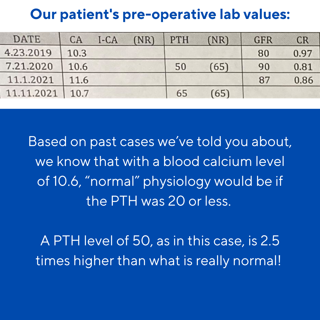
Preoperatively, his PTH was in the 50s, but his calcium levels were 10.5 and 10.7.

In roughly 20% of our patients, we see this phenomenon of "abnormally normal PTH" levels. This phenomenon can cause patients to suffer longer than necessary, since many providers will wait for the PTH level to increase above the "normal" range before recognizing their patient is suffering with primary hyperparathyroidism.

Meanwhile, the disease is wreaking havoc on the patient's body.

From our collective experience of over 60,000 cases, we know that it doesn't matter if your PTH is 50 or 150 – but it does matter what your PTH is doing in relation to your calcium level. And when your calcium level is 10.5, your PTH should be less than 20. The purpose of the parathyroid glands is to regulate the blood calcium level and recognize that when the calcium level is that high, PTH does not need to be released into the system to pull more calcium from the bones into the bloodstream.

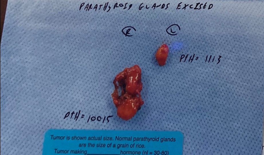
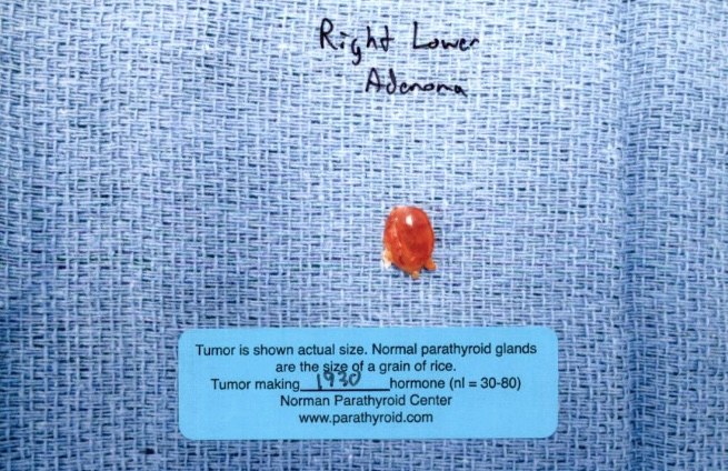
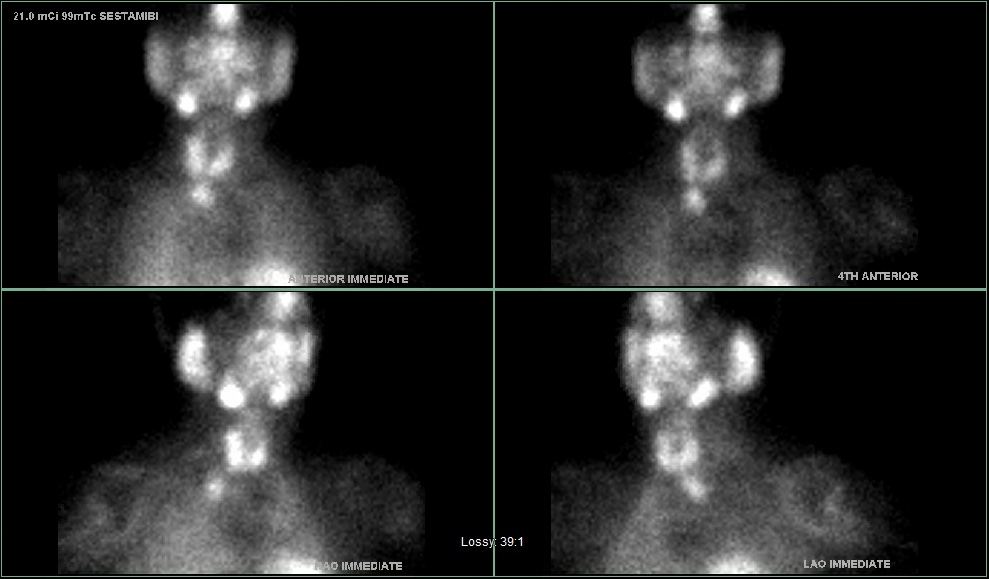
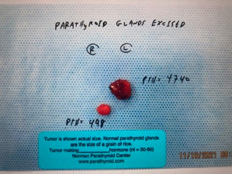
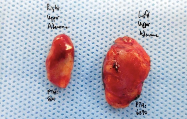
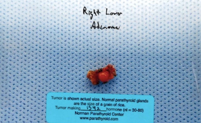
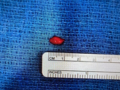
Intraoperatively, two parathyroid tumors were found. Normal parathyroid gland intraoperative PTH production is roughly 50 units, with his tumors producing 333 and 2267 units. His post-operative blood PTH was 15. This is a great example of a patient with "normal" pre-op PTH levels being cured with surgery.

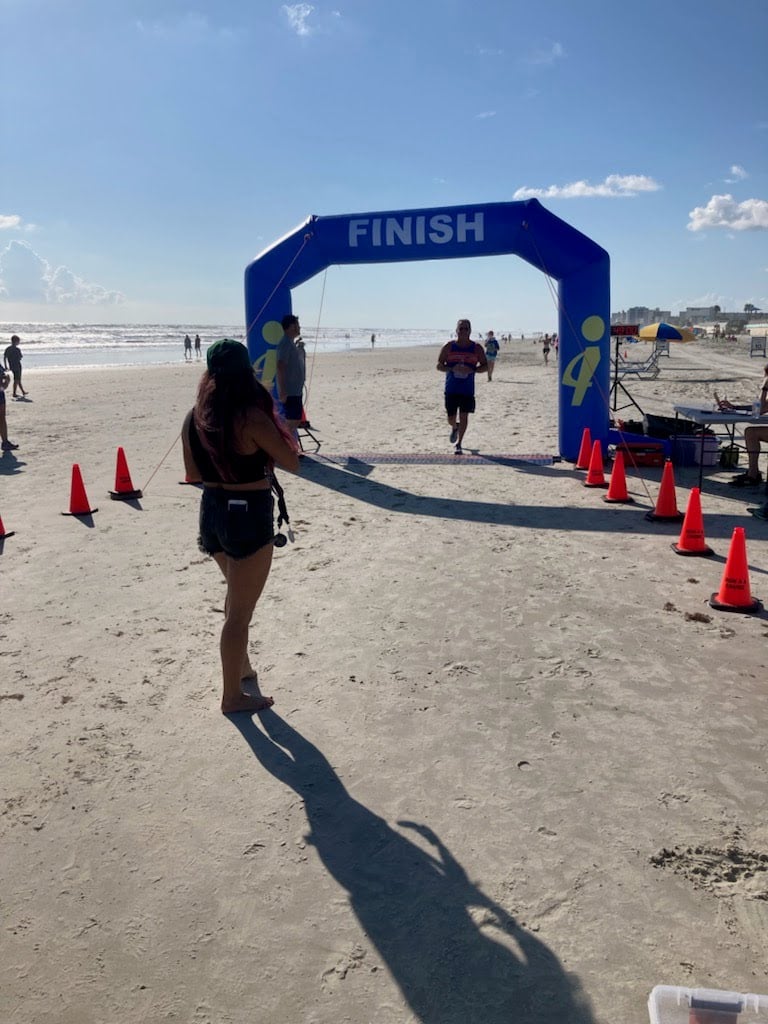
The evening before his surgery, he told me he had been unable to figure out a math problem for his current coursework toward another graduate degree. On the night of surgery, the problem made sense! Just three days after surgery, he sent me a picture of him crossing the finish line at his first race in years. He said it was his "couch to 5K" miracle.

We know there was no miracle, but rather good surgery allowing him to feel whole again.


Image

Interesting case of the week 10/15/2: Another parathyroid gland trying, and failing, to hide with Dr. Luke Watkins

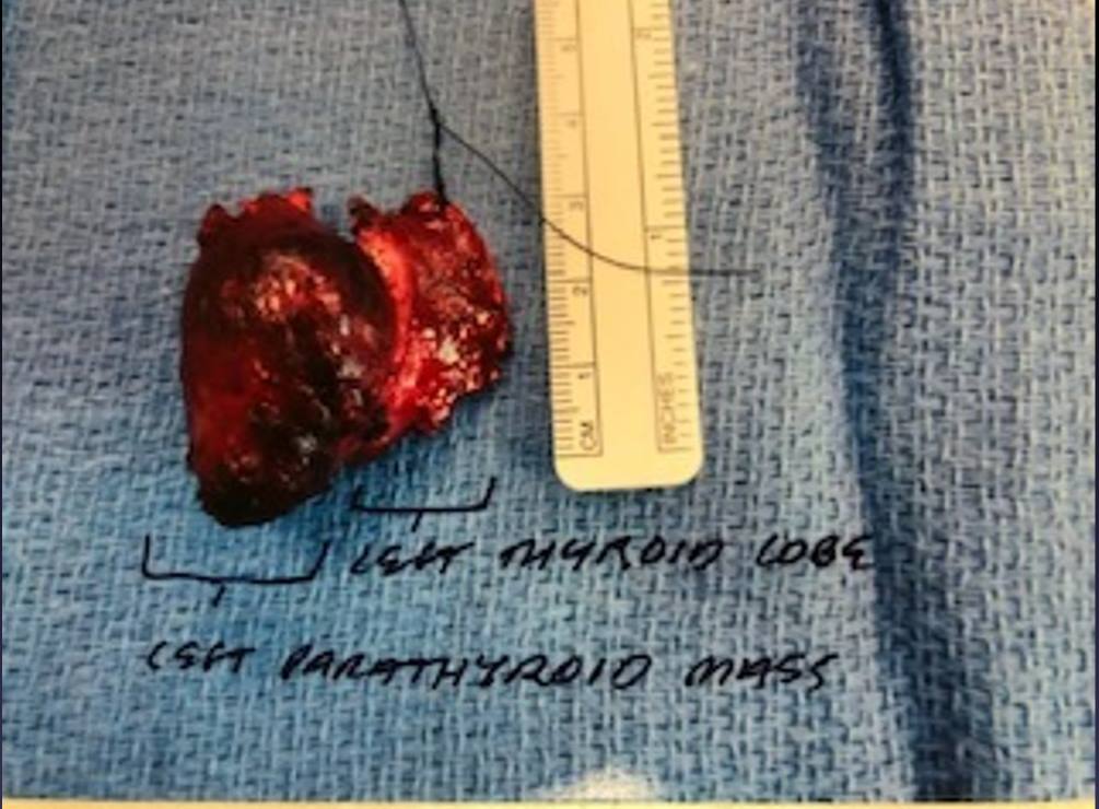
This week we're discussing a 56-year-old female who travelled across the country to see us for her parathyroid surgery. Her calcium level was 10.6 with a PTH of 81, clearly demonstrating primary hyperparathyroidism.

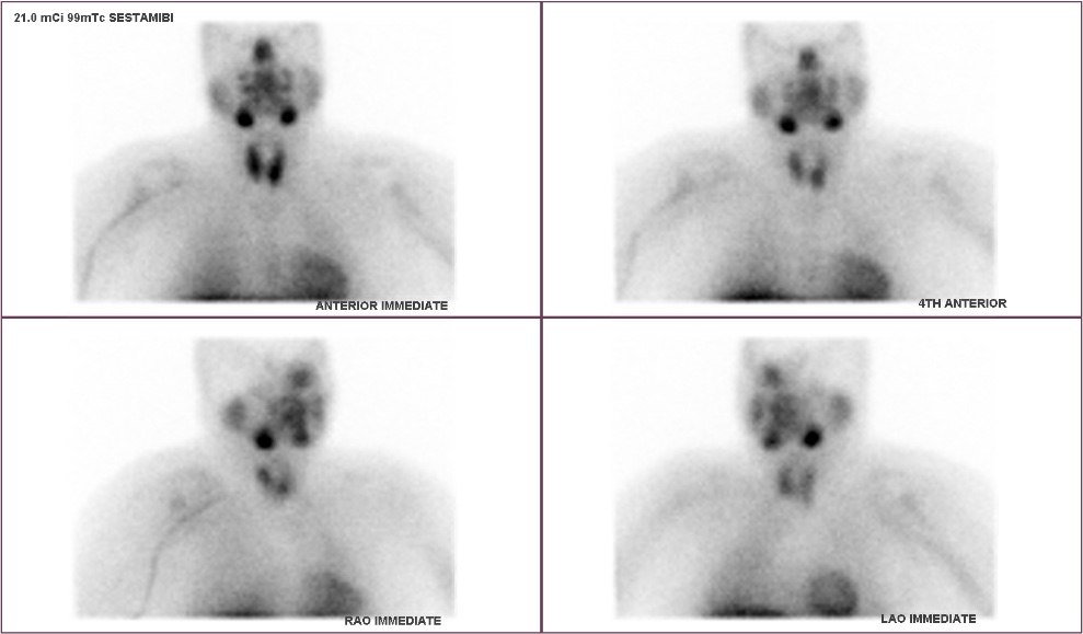
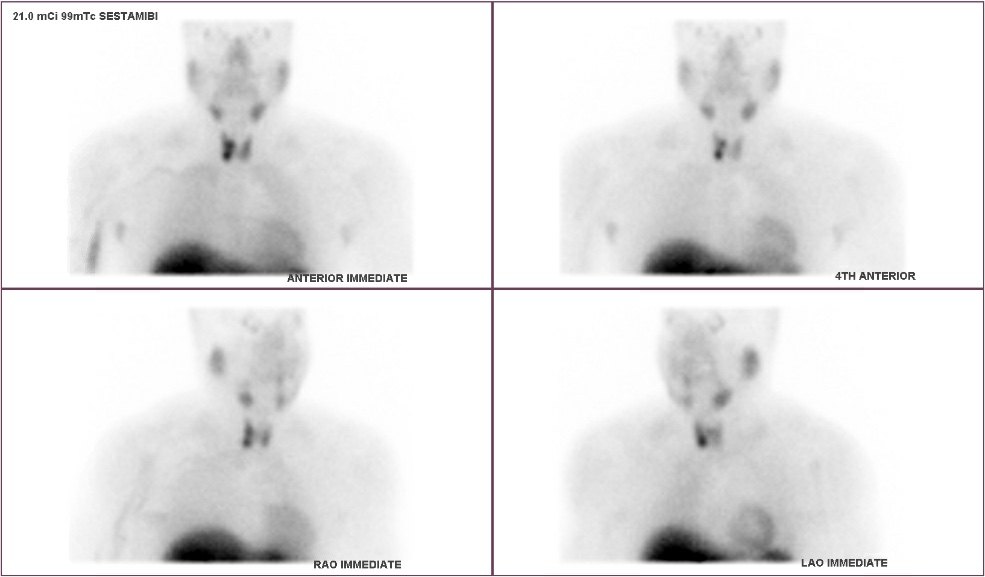
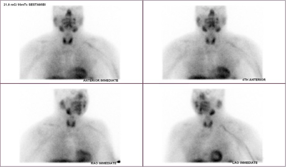
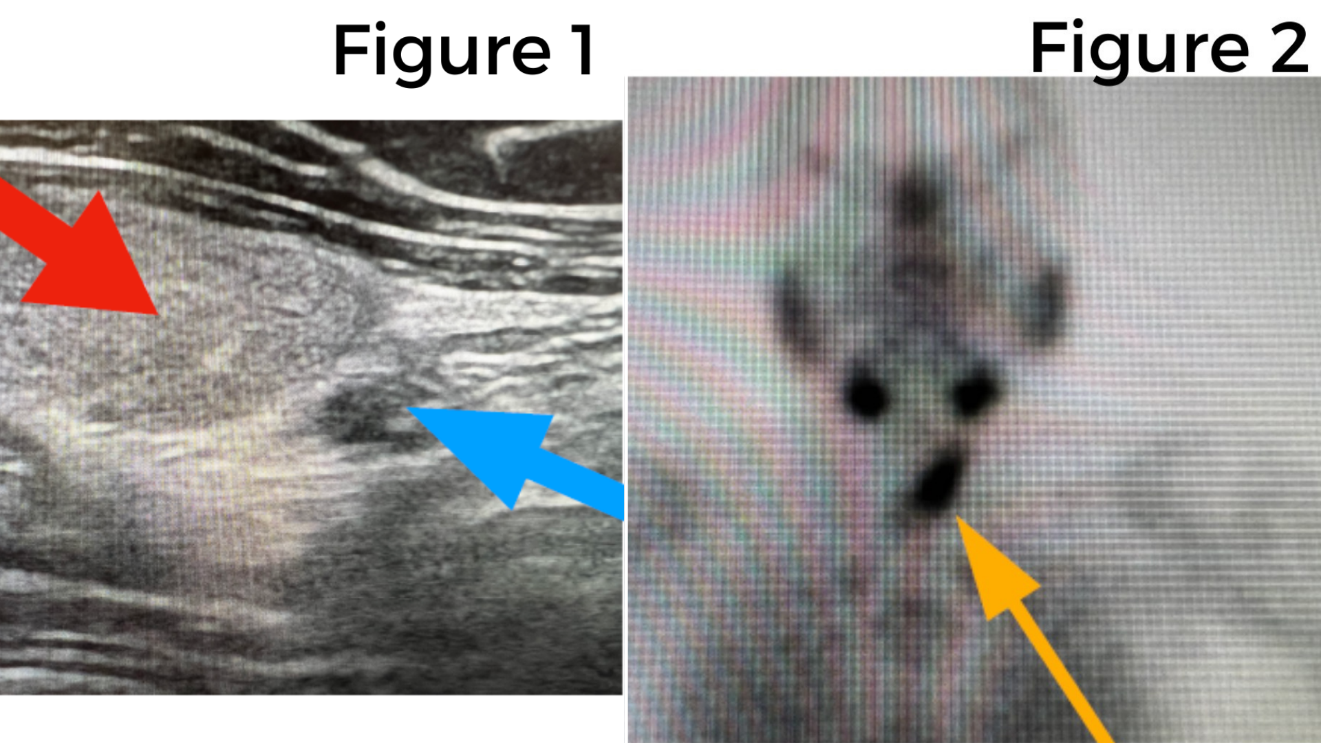
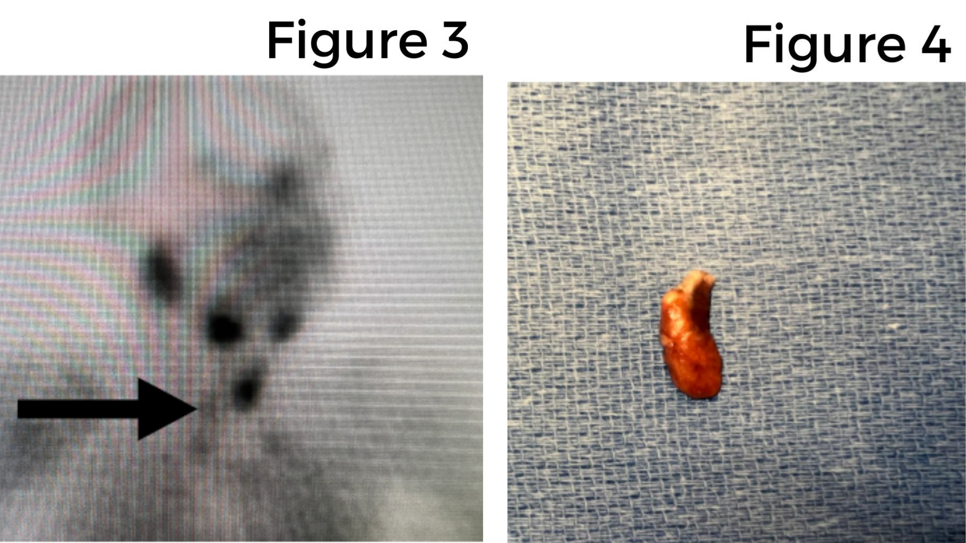
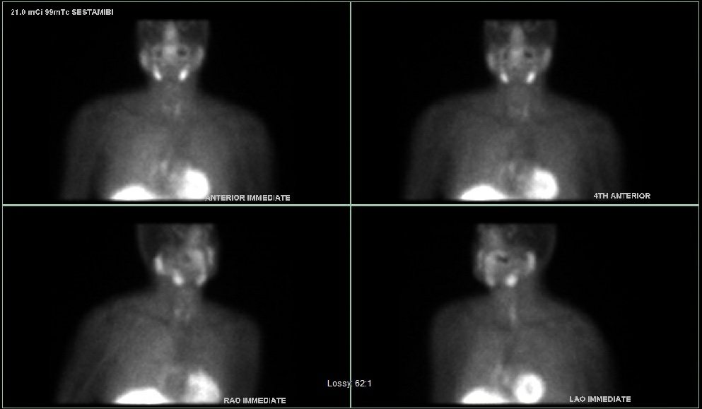
She had been taking lithium, which can cause hyperparathyroidism, and had worsening kidney function. She also had multiple symptoms, including fatigue, insomnia, irritability, and brain fog. Prior to being sent to us, she had a sestamibi scan and an ultrasound that did not note any parathyroid glands.

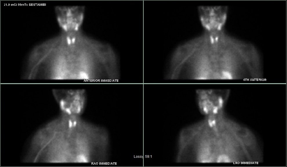
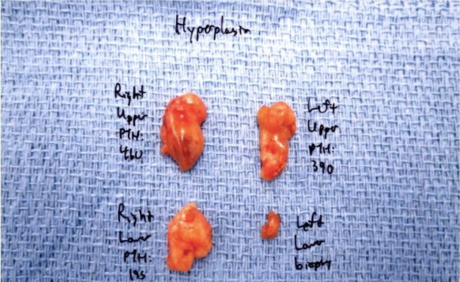
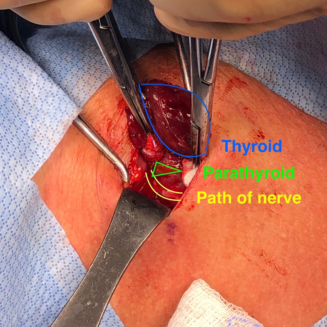
During her surgery, we quickly found three mildly enlarged, or hyperplastic, parathyroid glands. One gland was not in any of the usual places. But we know where these glands like to hide. After checking the more common locations, we separated part of the thyroid gland from its blood supply to fold the upper part down and look behind it. Her fourth gland was hiding between the thyroid and the windpipe, just millimeters away from the nerve for her voice.

Our knowledge of parathyroid anatomy and experience helped us find this gland, which was over-functioning. Without finding it, she would not have been cured.

The image below was taken at the time of surgery and is marked to show the thyroid gland being retracted in blue, the path to the nerve to the vocal cords between the yellow lines, and the parathyroid gland in the green triangle.


Image

Interesting case of the week 10/8/21: Parathyroid Carcinoma Presenting As Recurrent Primary Hyperparathyroidism with Dr. Jamie Mitchell

Today we’ll be delving into the case of 79-year-old woman diagnosed with primary hyperparathyroidism in 2015. She underwent surgery to remove a left lower parathyroid adenoma and her calcium and PTF levels normalized post-operatively.

In 2021, she was admitted to the hospital with dangerously high calcium levels of 14.0 mg/dl and PTH 508pg/ml.

She was re-evaluated for surgery, with the expectation of an additional adenoma on the right side of the neck which was not explored during her first operation.

Interestingly, her pre-operative sestamibi scan showed a large signal on the left, and her ultrasound went on to reveal a large mass that was compressing her left thyroid lobe. We removed the suspicious mass along with her left thyroid lobe.

Her pathology report revealed that the parathyroid tumor was consistent with parathyroid carcinoma.

Parathyroid carcinoma is very rare, accounting for less than 1% of cases of hyperparathyroidism. If diagnosis is known prior to surgery, the standard of care is to remove the tumor along with the thyroid lobe. Luckily, that’s exactly what was done in this case.

This patient is doing well and her post-operative care includes ongoing surveillance for recurrence with labs and imaging.

Interesting case of the week: Parathyroid Carcinoma Presenting As Recurrent Primary Hyperparathyroidism with Dr. Jamie Mitchell 2